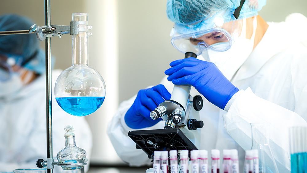
Researchers to 3D-print living human pancreas for diabetes research
03/10/2021 / By Virgilio Marin

An international team of researchers will be 3D printing live human pancreas models as part of a research initiative called the “European ENLIGHT Project.” The researchers plan to use the “bioprinted” models to improve the testing of diabetes medications and eventually treatments for other diseases like cancer.
The European Union‘s Horizon 2020 Innovation Fund awarded the project a four-year grant totaling 3.6 million euros ($4.3 million). Swiss tech company Readily3D, the project’s official bioprinter manufacturer, will be lending its contactless tomographic illumination technology, Tomolite 3D.
3D printing the human pancreas
The researchers aim to print its first set of working pancreas models within three years. To hit that target, they first need to adapt Tomolite 3D for the fabrication of pancreatic tissues.
If all goes according to plan, the final product will be able to print centimeter-scale tissue structures on a layer-by-layer basis in less than 30 seconds – virtually at “lightning speeds” compared to conventional 3D printers which take hours to achieve the same thing. The printing speed is especially important as the survival rate of living cells dramatically decreases as print times increase.
“With its rapid build speed, low light dose and sterile build environment, our tomographic bioprinter will open up previously inaccessible applications in biofabrication,” said Readily 3D’s chief technical officer Paul Delrot.
Once the bioprinter is able to create living pancreas models, the researchers will then add signaling molecules to the bioprinted tissues to determine how pancreatic cells behave under external stimulation. This will essentially recreate the function of the cells in a living system, which will enable researchers to use the structures as a test pancreas for diabetes research without resorting to animal testing.
Project coordinator Riccardo Levato, a biofabrication researcher at the University Medical Center Utrecht in the Netherlands, which will co-host the printing of the models, said that introducing cells from a diabetes patient will allow researchers to recreate diseased tissue and determine which candidate medication works best.
“This spares patients a long search with unpleasant side effects, saves on treatment costs and leads to the best available care for individual patients,” Levato said. (Related: 3D printers may revolutionize more than just consumer products – Scientists can now print human ’tissue’ and functional artificial ears.)
The researchers chose to focus on diabetes because the condition is one of the most common chronic diseases among children. Additionally, the development of new diabetic treatments has been falling behind.
But the bioprinting technique can eventually be scaled up for other diseases such as cancer. According to Levato, the pancreas models will serve as “proof of principle.” If Levato and his team manage to make a living model of the pancreas and test diabetes medication with it, then that would prove that their bioprinting technique is effective.
“Then we can use those techniques much more widely. In principle, you can make living models of all types of tissue with it,” Levato noted.
Making a human heart from a seaweed-based material
Meanwhile, researchers from Carnegie Mellon University 3D printed a lifelike heart model using a seaweed-based material called alginate. In a paper published in the journal ACS Biomaterials Science & Engineering, the researchers described how they used a 3D printing technique called Freeform Reversible Embedding of Suspended Hydrogels (FRESH) to reproduce a life-size organ.
This technique involves 3D printing soft biomaterials within a gelatin bath to support delicate structures that would otherwise fall apart in the air. It’s able to replicate the feel of natural tissues but its use had been limited to making small objects prior to the study.
The researchers then tried to create a human heart to adapt the technique for bigger organs. To that end, they used alginate because it has similar mechanical properties as heart tissue. This tensile material remains intact even when stretched, which means that surgeons-in-training can practice stitching up a heart model made from the material.
Using a modified FRESH 3D printer and heart scans from a patient, the researchers printed a full-size adult human heart and a section of the coronary artery that could be filled with fake blood. The heart model was structurally accurate, reproducible and intact even when taken outside of the gelatin bath.
The researchers concluded that FRESH is a viable technique for printing realistic organ models that can be used to train future surgeons.
Learn more about amazing medical breakthroughs at FutureScienceNews.com.
Sources include:
Tagged Under: 3D printing, biofabrication, bioprinting, biotechnology, breakthrough, diabetes, future science, future tech, goodtech, innovation, medical tech, research, science and technology
RECENT NEWS & ARTICLES
COPYRIGHT © 2018 BREAKTHROUGH.NEWS
All content posted on this site is protected under Free Speech. Breakthrough.news is not responsible for content written by contributing authors. The information on this site is provided for educational and entertainment purposes only. It is not intended as a substitute for professional advice of any kind. Breakthrough.news assumes no responsibility for the use or misuse of this material. All trademarks, registered trademarks and service marks mentioned on this site are the property of their respective owners.

